Sección científica.Primer premio = First Award Artistic Section.
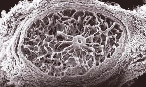
Región retrolaminar de un nervio óptico humano.
Imagen obtenida con microscopía de barrido (SEM) en una preparación de nervio previamente digerida con tripsina para la eliminación de todo el tejido nervioso. Con esta técnica se pone de manifiesto todo el entramado de tejido conectivo que forma la tabicación de la región retrolaminar y que se extiende en continuidad desde la envuelta de un vaso central hasta el tabique pial periférico.
Human optic nerve: retrolaminar region
Image obtained using scanning electron microscopy. The optic nerve was prepared by digestion with trypsin to remove the nervous tissue and to reveal the connective tissue. This technique reveals the structure of the connective tissue that makes up the wall of the retrolaminar region, extending from the adventitia of the central vessel to the peripheral pia mater.
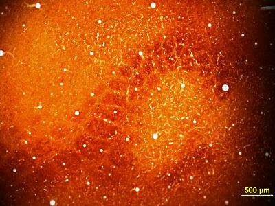
55. Coral de colágeno


