Inicio Biblioteca Complutense Catálogo Cisne Colección Digital Complutense

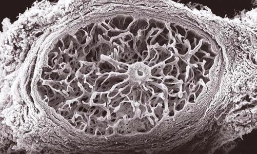
55. Coral de colágeno

Rosa de Hoz Montañana 30 de Mayo de 2011 a las 17:34 h

Ver texto

Sección científica.Primer premio = First Award Artistic Section.

Región retrolaminar de un nervio óptico humano.

Imagen obtenida con microscopía de barrido (SEM) en una preparación de nervio previamente digerida con tripsina para la eliminación de todo el tejido nervioso. Con esta técnica se pone de manifiesto todo el entramado de tejido conectivo que forma la tabicación de la región retrolaminar y que se extiende en continuidad desde la envuelta de un vaso central hasta el tabique pial periférico.

Human optic nerve: retrolaminar region

Image obtained using scanning electron microscopy.  The optic nerve was prepared by digestion with trypsin to remove the nervous tissue and to reveal the connective tissue. This technique reveals the structure of the connective tissue that makes up the wall of the retrolaminar region, extending from the adventitia of the central vessel to the peripheral pia mater.

  55. Coral de colágeno

20. Fento

Manuel Solalleiro 30 de Mayo de 2011 a las 17:25 h

Sección científica. Segundo Premio = Scientific Section. 2nd award


A fern in bud revealing how Nature creates her own geometrical growth patterns.


Brote de helecho que muestra cómo la naturaleza toma estructuras geométricas en fases de crecimiento.

  20. Fento

27. Palafitos Chiloe

Camila Vásques Navarro 30 de Mayo de 2011 a las 17:22 h

Sección científica. Tercer premio = Scientific Section. 3rd award.

Double vision in a multicoloured world

This photograph was taken last year while on a visit to Chile, the country in which I was born. The photograph shows the reflection of the coloured houses in the fishing villageof Palafitos, in the region of Chiloé to the south of Chile.

I consider it to be a scientific photograph because the theme of reflection, as may be appreciated in the sea, formed part of my studies. The symmetry is well defined and the optical effect is well achieved by the clarity with which the sea is portrayed. The colours have been enhanced to give the photograph an extra beauty.

[Seguir leyendo] 27. Palafitos Chiloe

53. El árbol de los huevos = The egg tree

Juan J. Salazar Corral 30 de Mayo de 2011 a las 17:17 h

Sección científica. Áccesit

A stereo image obtained using a Technival Carl Zeiss Jena stereomicroscope, which shows part of the ocular vasculature in two diaphanized eyeballs.

The diaphanization technique is based on the principle that a tissue refracts the least amount of light and reaches its maximum transparency when it is surrounded and embedded in a substance whose refractive index equals the average refractive index of the tissue. If the blood vessels have previously been infused with a coloured resin, the ocular vasculature can be studied.

[Seguir leyendo] 53. El árbol de los huevos = The egg tree

85. Lateral line organs

Gary Baker 30 de Mayo de 2011 a las 17:05 h

Scientific Commended

Distribution of melanopsin-positive immuno-histochemical staining in a wholemount of skin from a part of the dorsal surface of an adult Xenopus  laevis. The image shows part of a lateral line stitch with three immuno-positive lateral line organs surrounded by several small circular immuno-positive dermal melanophores. Two separate compartments of dense immunopositive staining are evident in each organ with fine immuno-positive processes emanating away from the centre.

The tissue was incubated in a 1:2000 dilution of a polyclonal antibody to melanopsin raised in rabbit against Xenopus dermal melanophores (CERN972; de Grip). The second antibody (biotinylated anti-rabbit - Vector Labs) was revealed using diaminobenzidine. Control incubations in the absence of either the primary or the second antibody resulted in tissue devoid of immunostaining.

  85. Lateral line organs

81. Spectrum

Luis Díaz-Santana 30 de Mayo de 2011 a las 17:02 h

Scientific Commended. 

This picture is an allegory of the electromagnetic spectrum, where only the visible part of the spectrum is colour. The waveform image is itself an allegory of the Huygens' Principle. The pencil is one of the most powerful tools of the scientist: creativity.


Esta fotografía es una alegoría del espectro electromagnético, donde únicamente la parte visible del espectro tiene color. La imagen en forma de onda es a su vez una alegoría del principio de Huygens. Los lápices representa una de las herramientas más poderosas del científico: la creatividad.

  81. Spectrum


Universidad Complutense de Madrid - Ciudad Universitaria - 28040 Madrid - Tel. +34 914520400
[Información - Sugerencias]